
Neoplasms

This circumscribed reddish-yellow firm neoplasm beneath the dura next to the falx is a meningioma. The superior parasagittal location is quite common.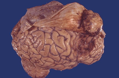 |

Here is a resected meningioma. These are relatively easy for the neurosurgeon to remove.

At high magnification, this meningioma has plump pink cells. A small amount of brown granular hemosiderin is present. Meningiomas may also have psammomma bodies.

Here is an ependymoma arising from the ependymal lining of the fourth ventricle above the brainstem and bulging toward the cerebellum. Ependymomas are benign histologically

This horizontal (CT scan) section of the brain reveals a large ependymoma of the fourth ventricle. |
The microscopic appearance of an ependymoma reveals a rosette pattern with the cells arranged about a central vascular space. It is histologically benign. |

Here is the microscopic appearance of a medulloblastoma with small round blue cells. |

The mass lesion here is arising in the acoustic (eighth cranial) nerve at the cerebellopontine angle. This is a schwannoma. Patients may present with hearing loss. These benign neoplasms can be removed.

This sagittal section of brain demonstrates a large brainstem glioma. Most gliomas are astrocytomas. This astrocytoma demonstrates increased cellularity and pleomorphism, as compared to normal brain. Note the very pleomorphic cell in the center. |

This glioblastoma multiforme (GBM) demonstrates marked cellularity with marked hyperchromatism and pleomorphism. Note the prominent vascularity as well as the area of necrosis at the left with neoplastic cells palisading around it.

| At the left is seen a metastasis from a lung carcinoma. Metastases most often appear at the border of the grey and white matter in the distribution of the middle cerebral artery, as in this case, because that is where the blood flow (vascular distribution) is most likely to take metastases. |