Myocarditis
The epicardial surface of the heart is smooth and glistening, but there are small scattered pinpoint yellowish microabscesses
The microscopic appearance of a microabscess is shown here. The center consists of blue bacterial colonies and is surrounded by acute inflammatory cells.
Microscopically, acute rheumatic carditis is marked by a peculiar form of granulomatous inflammation with so-called "Aschoff nodules" seen best in myocardium. These are centered in interstitium around vessels as shown here. The myocarditis may be severe enough to cause congestive heart failure
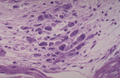
Here is an Aschoff nodule at high magnification. The most characteristic component is the Aschoff giant cell. Several appear here as large cells with two or more nuclei that have prominent nucleoli. Scattered inflammatory cells accompany them and can be mononuclears or occasionally neutrophils.
The interstitial lymphocytic infiltrates shown here are characteristic for a viral myocarditis, which is probably the most common type of myocarditis. Many of these cases are probably subclinical. Some may be a cause for sudden death in young persons. There is usually little necrosis. The most common viral agent is Coxsackie B.
In time, chronic rheumatic valvulitis may develop by organization of the acute endocardial inflammation along with fibrosis, as shown here affecting the mitral valve. Note the shortened and thickened chordae tendineae.
________________________________________________________________-
Neoplasia
This two year old child died suddenly. At autopsy, a large firm, white tumor mass was found filling much of the left ventricle. This is a cardiac rhabdomyoma. Such primary tumors of the heart are rare.
This high power microscopic appearance of cardiac myxoma shows minimal cellularity. Only scattered spindle cells with scant pink cytoplasm are present in a loose myxoid stroma.
Primary tumors of the heart are uncommon. Metastases to the heart are more common, but rare overall (only about 5 to 10% of all malignancies have cardiac metastases). Seen over the surface of the epicardium are pale white-tan nodules of metastatic tumor. Metastases may lead to a hemorrhagic pericarditis.
The neoplasm with the greatest propensity to metastasize to heart is melanoma. The metastatic melanoma is seen here to be infiltrating into the myocardium. At the arrow can be seen some brown-black pigment characteristic of melanoma.
The left atrium has been opened to reveal the most common primary cardiac neoplasm--an atrial myxoma. These benign masses are most often attached to the atrial wall, but can arise on a valve or in a ventricle. They can produce a "ball valve" effect by intermittently occluding the atrioventricular valve orifice. Embolization of fragments of tumor may also occur. Myxomas are easily diagnosed by echocardiography.


